Image Credits
Articles on APO|APE News that do not carry a photograph of their own are illustrated with real microscopy, histology and molecular-structure imagery drawn from public-domain and Creative Commons sources. Every image is reproduced in the publication's colour palette: the original is converted to a single-tone rendering in purple and magenta, cropped to a 16:9 frame, and in some cases inverted so a structure drawn on white sits on a dark ground. No scientific content is added. The originals, their creators and their licenses are listed below; follow the source link to see each image as published.
Images marked CC BY or CC BY-SA are used under the Creative Commons license named; our tinted versions of the CC BY-SA images are available under the same license on request. Public-domain and CC0 images carry no license condition, and are credited here as a courtesy to their creators.
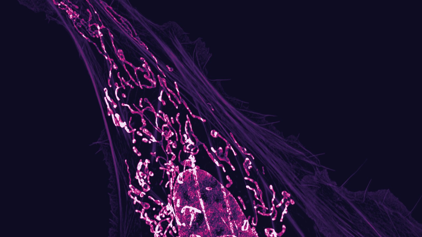
Osteosarcoma cell: nucleus, mitochondria and actin filamentsDylan Burnette and Jennifer Lippincott-Schwartz, National Institutes of Health
Public domain ·
Source 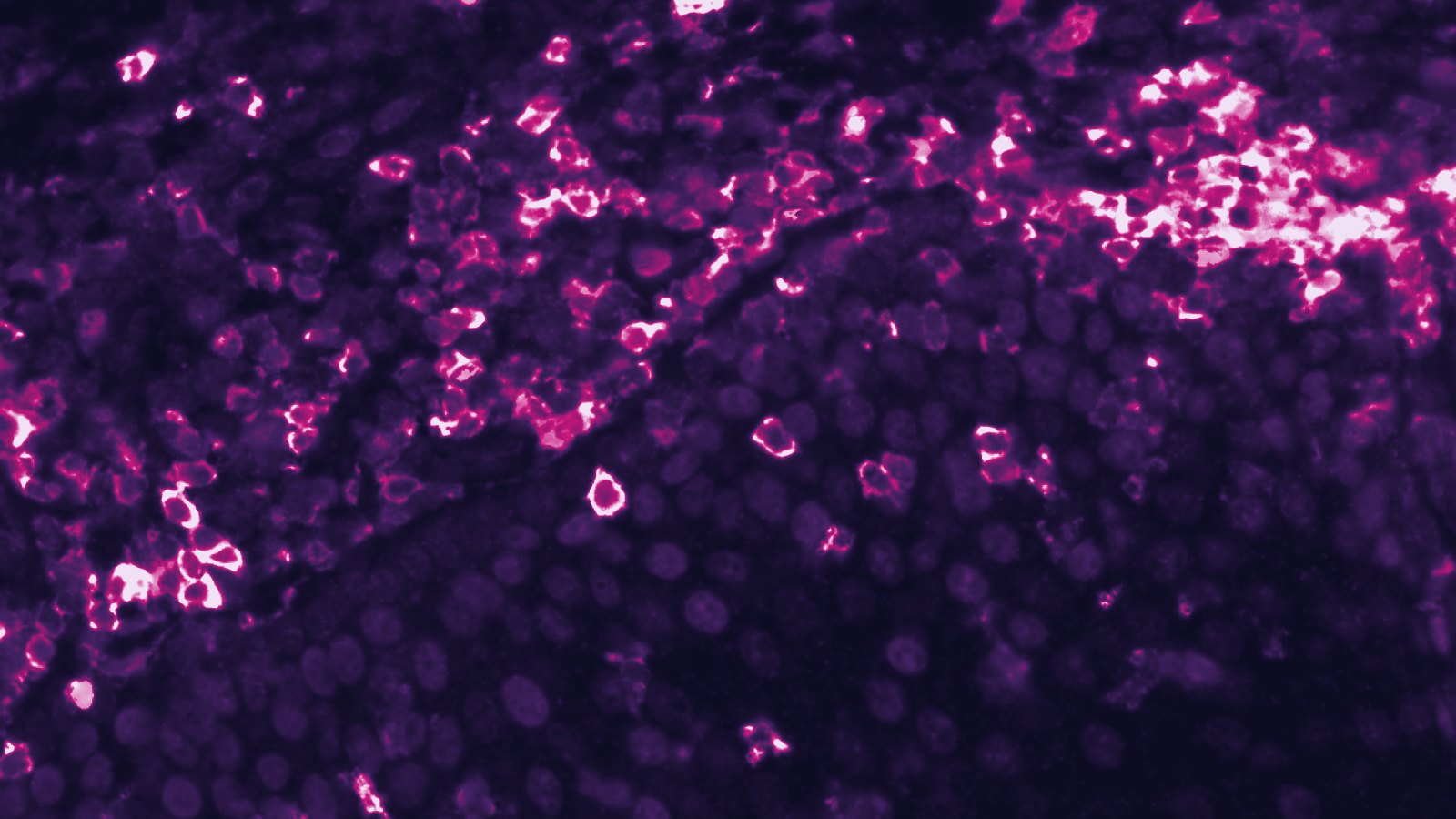
T cells infiltrating a squamous cell carcinomaNational Institutes of Health, via Wikimedia Commons
Public domain ·
Source 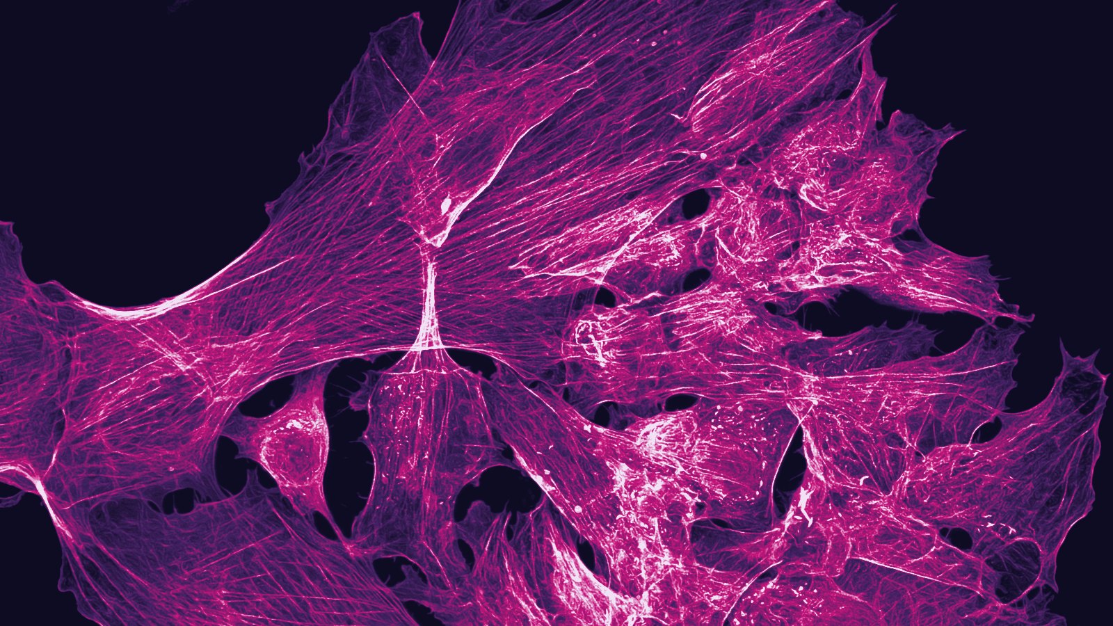
Osteosarcoma cells stained for tubulin, actin and nucleiHoward Vindin, via Wikimedia Commons
CC BY 4.0 ·
Source 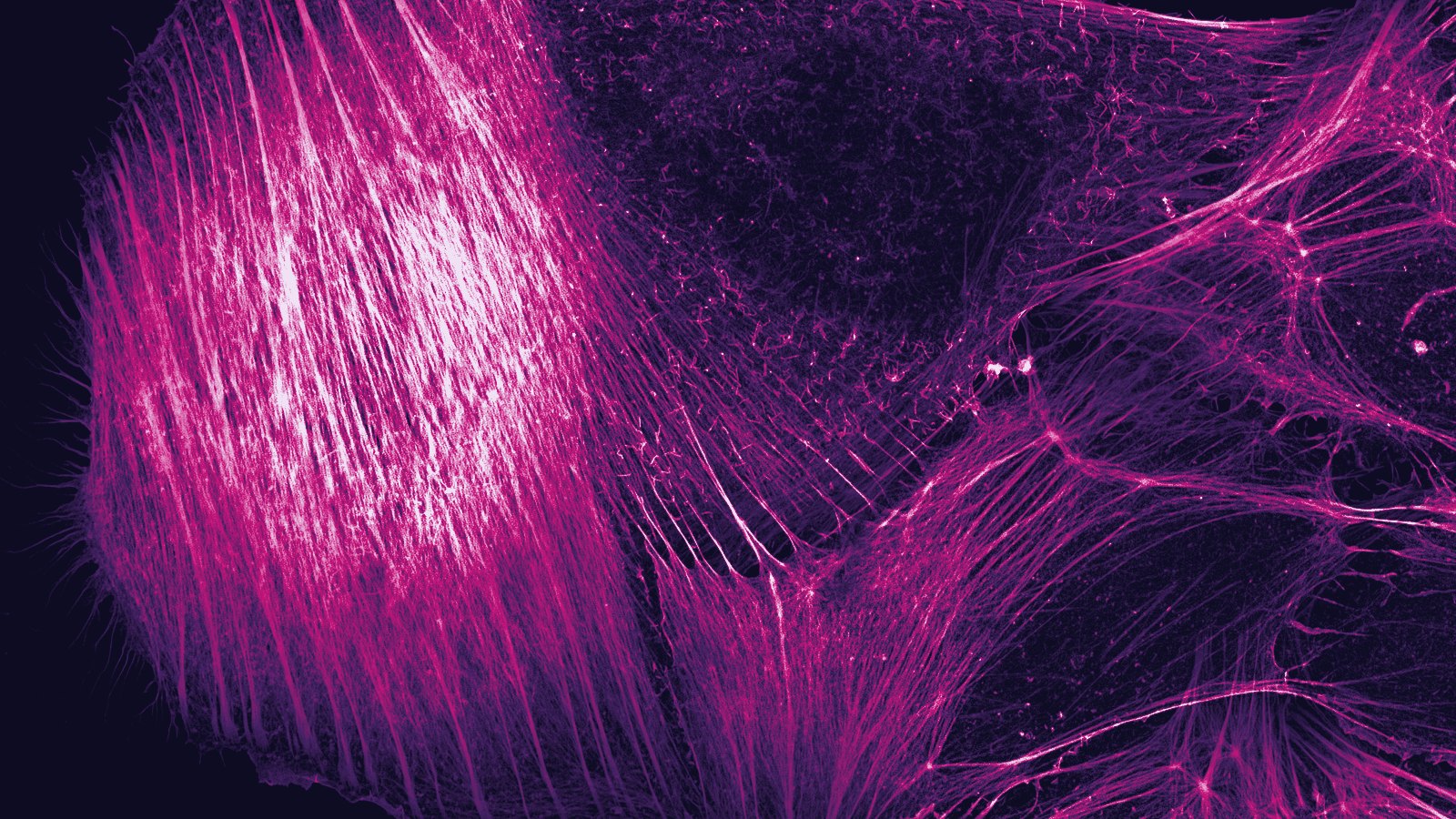
Osteosarcoma cell, depth-coded actin filamentsHoward Vindin, via Wikimedia Commons
CC BY-SA 4.0 ·
Source 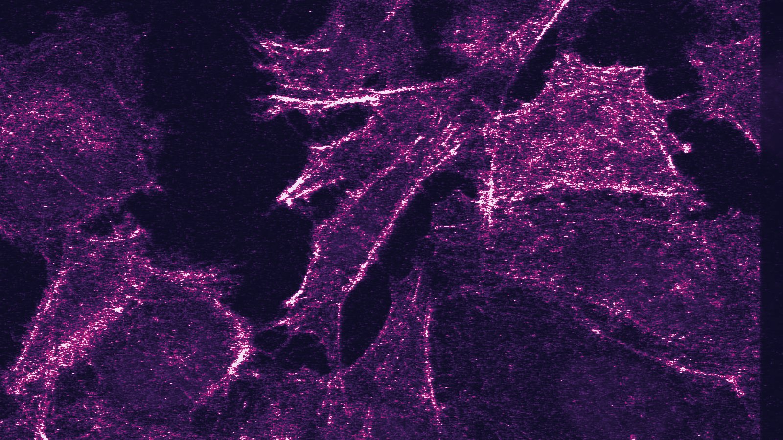
PC3 prostate cancer cells, confocal microscopy (actin and nuclei)Griersonj5, via Wikimedia Commons
CC BY-SA 4.0 ·
Source 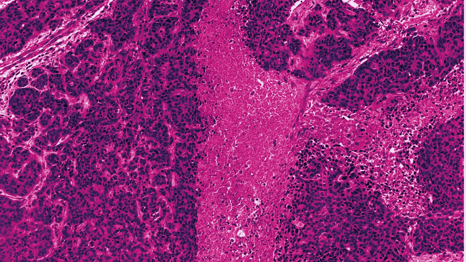
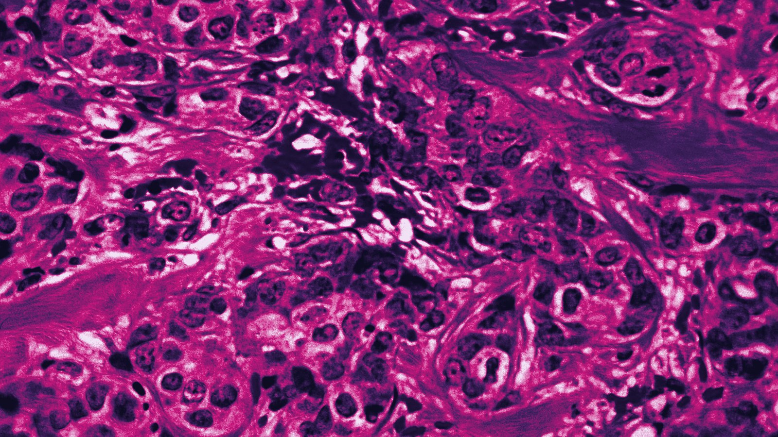
Basal-like breast cancer, H&EMikael Häggström, M.D., via Wikimedia Commons
CC0 1.0 ·
Source 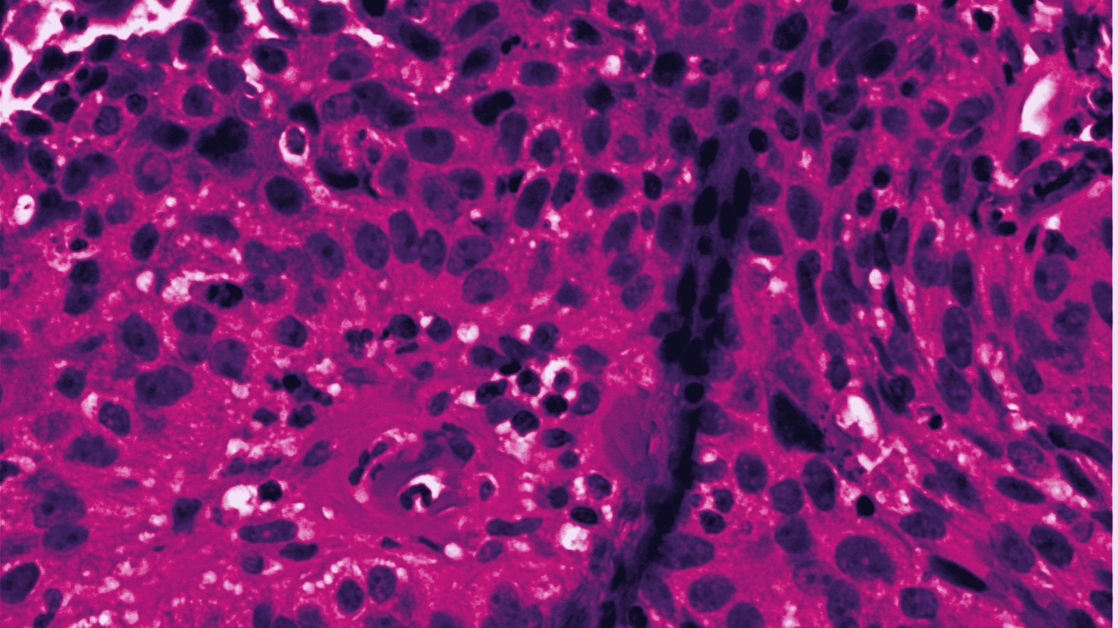
Invasive cervical squamous cell carcinoma, H&EMikael Häggström, M.D., via Wikimedia Commons
CC0 1.0 ·
Source 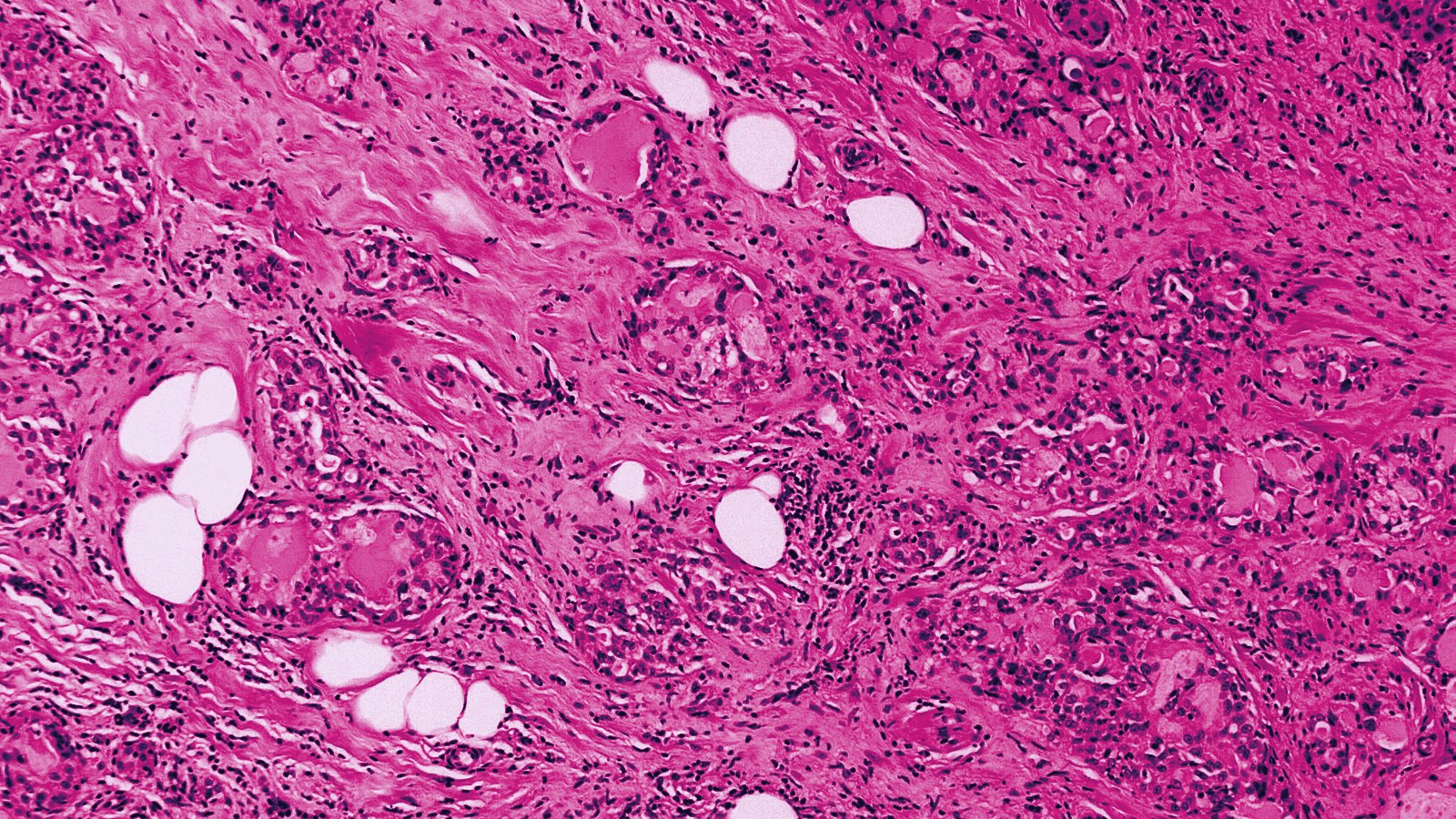
Secretory carcinoma, H&E, low magnificationMikael Häggström, M.D., via Wikimedia Commons
CC0 1.0 ·
Source 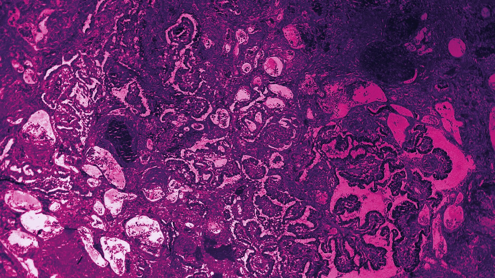
Encapsulated papillary carcinoma of the breast, H&EEd Uthman, M.D., via Wikimedia Commons
CC BY 2.0 ·
Source 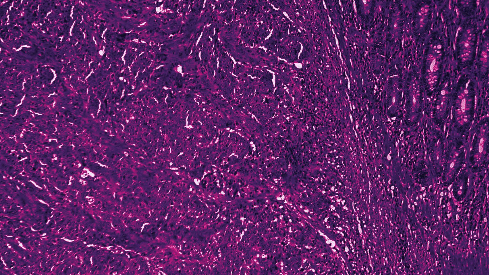
Medullary carcinoma of the colon, H&EEd Uthman, M.D., via Wikimedia Commons
CC BY 2.0 ·
Source 
HER2 receptor bound by trastuzumab (molecular painting)David S. Goodsell, RCSB Protein Data Bank, Molecule of the Month 268
CC BY 4.0 ·
Source 
PD-1 bound to PD-L1 and PD-L2 (molecular painting)David S. Goodsell, RCSB Protein Data Bank, Molecule of the Month 204
CC BY 4.0 ·
Source 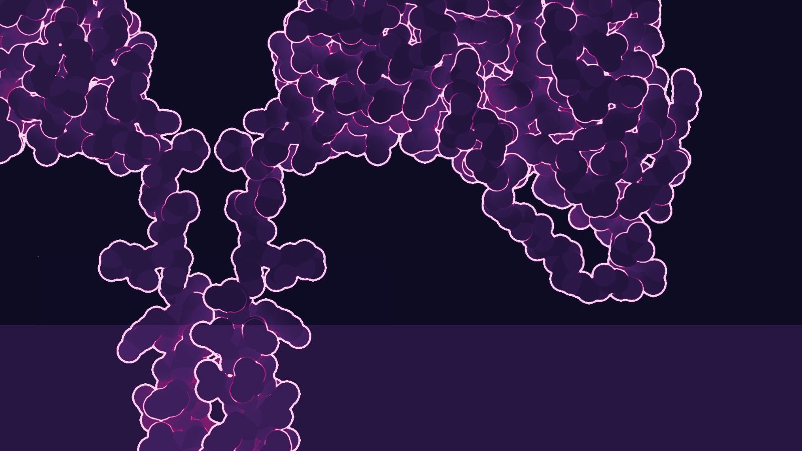
Chimeric antigen receptor (molecular painting)David S. Goodsell, RCSB Protein Data Bank, Molecule of the Month 214
CC BY 4.0 ·
Source 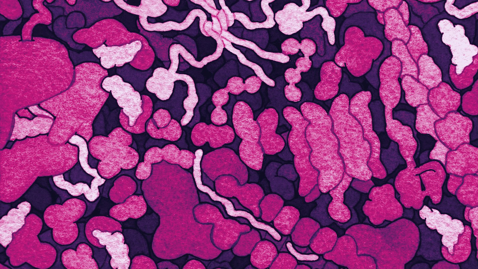
MDM2 and p53 inside a cell (molecular painting)David S. Goodsell, RCSB Protein Data Bank, Molecule of the Month 234
CC BY 4.0 ·
Source 
c-Abl kinase, the imatinib target (molecular painting)David S. Goodsell, RCSB Protein Data Bank, Molecule of the Month 283
CC BY 4.0 ·
Source 
VEGF and angiogenesis (molecular painting)David S. Goodsell, RCSB Protein Data Bank, Molecule of the Month 267
CC BY 4.0 ·
Source 
Blood smear showing acute lymphoblastic leukaemiaWellcome Collection, via Wikimedia Commons
CC BY 4.0 ·
Source 
Red blood cell, platelet and white blood cell, scanning electron micrographBruce Wetzel and Harry Schaefer, National Cancer Institute, via Wikimedia Commons
Public domain ·
Source Corrections
If you created one of these images and the credit is wrong or incomplete, write to [CONTACT EMAIL PLACEHOLDER] and it will be corrected.



.jpg)
.jpg)

