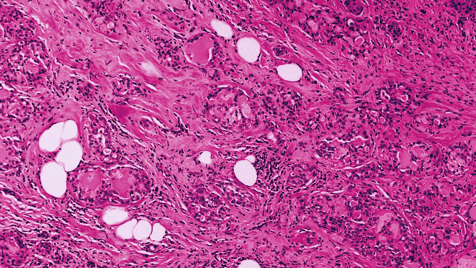
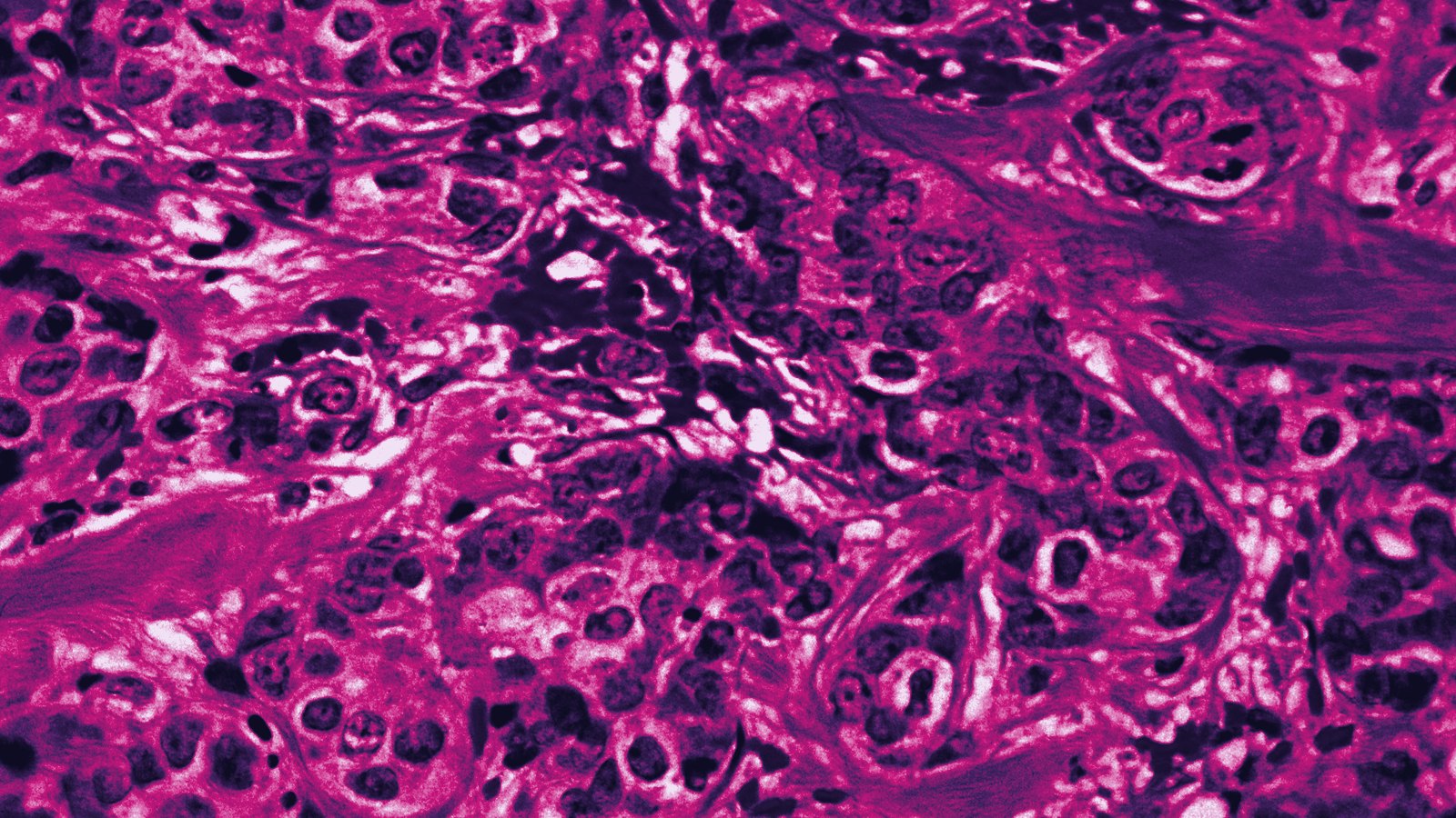
Atypical glandular cells appeared in 141 of 128,022 cervical cytology specimens at a single Istanbul gynecologic oncology center, and among the 119 women who reached histologic follow-up, 28 had neoplastic disease. Endometrial carcinoma made up the largest share of those outcomes. Age, dual p16 and Ki-67 positivity, and loss of nuclear polarity survived multivariable adjustment as independent predictors.
- Frequency: 141 AGC diagnoses among 128,022 cervical cytology specimens, 0.11 percent.
- Window: cases accessioned January 2013 through December 2023 at Kartal Lutfi Kirdar City Hospital, Istanbul; 119 patients analyzed after exclusions.
- Outcome: neoplastic pathology in 28 patients, 23.5 percent.
- Distribution: endometrial carcinoma 39.3 percent of the 28, cervical neoplasia 35.7 percent, metastatic malignancy 17.9 percent, ovarian high-grade serous carcinoma 7.1 percent.
- Independent predictors: loss of polarity OR 16.873 (95% CI 4.012 to 70.953), p16 and Ki-67 positivity OR 7.321 (1.828 to 29.318), age OR 1.141 per year (1.072 to 1.214).
- Model: area under the receiver operating characteristic curve 0.918.
Kartal Lutfi Kirdar City Hospital, 2013 to 2023
Cervical cytology specimens accessioned at Kartal Lutfi Kirdar City Hospital in Istanbul between January 2013 and December 2023 yielded 141 diagnoses of atypical glandular cells, a rate of 0.11 percent. The paper calls itself a 10-year retrospective cohort study while reporting an 11-calendar-year case window. Exclusions, chiefly the requirement that histopathologic follow-up land within 12 months, left 119 patients. Neoplastic pathology turned up in 28 of them, 23.5 percent. Slides were rereviewed against the 2014 Bethesda System before analysis.
Those 28 cancers did not all originate in the cervix. Endometrial carcinoma accounted for 39.3 percent, cervical neoplasia for 35.7 percent, metastatic malignancy for 17.9 percent and ovarian high-grade serous carcinoma for 7.1 percent. A quarter of the neoplastic outcomes were therefore ovarian or metastatic, arising outside the field a colposcopist can see.
Loss of Polarity Carried the Largest Odds Ratio
Univariable analysis linked neoplastic outcome to increasing age, postmenopausal status, p16 and Ki-67 positivity, an increased nuclear-to-cytoplasmic ratio, nuclear enlargement, and loss of polarity. Three survived adjustment. Loss of polarity produced an odds ratio of 16.873 (95 percent CI 4.012 to 70.953, P less than 0.001), dual p16 and Ki-67 positivity 7.321 (1.828 to 29.318, P = 0.005), and each additional year of age 1.141 (1.072 to 1.214, P less than 0.001). The fitted model reached an area under the curve of 0.918. The intervals around the two categorical predictors run wide, which follows from fitting three terms to 28 events.
Esra Keles of the gynecologic oncology department and four co-authors, including pathologists Mervenur Sahin and Sibel Cetinkaya, conclude that "integration of these parameters with thorough clinical history may improve risk stratification and clinical management of patients with AGC." Corresponding author Sahra Sultan Kara works in obstetrics and gynecology at the same hospital, and Pinar Birol Ilter is at Kutahya City Hospital.
Where the p16 and Ki-67 Result Came From
The methods place the immunostaining on follow-up tissue specimens, not on the cytology preparation, which limits how the finding can be used at sign-out. A cytopathologist holding an AGC smear and no biopsy does not yet have the second-strongest predictor in the model. The two variables available at the moment the cytology is signed, the patient's age and loss of polarity on the slide, are also the ones that bracket the model's weighting, and neither costs anything to record.
The Abstract Omits the Bethesda Subcategories
No PubMed Central copy of the paper has been deposited, and Elsevier sells the rest. The published abstract and the affiliations returned by PubMed and Crossref were what remained. Four items a cytology service would want are missing from it. The 141 cases are not broken out by Bethesda subcategory, so the split between atypical glandular cells not otherwise specified, atypical glandular cells favor neoplastic, and endocervical adenocarcinoma in situ stays unknown. No immunohistochemistry beyond p16 and Ki-67 is reported, which leaves out ER, PAX8, CEA and vimentin, and no antibody clone or staining platform is named either. The analysis is multivariable logistic regression and receiver operating characteristic curves throughout, with no machine learning in the methods.
Why This Matters to the APO|APE Reader
Twenty-two women who received an AGC report never produced histology inside the study's follow-up window and were dropped from the analysis, so the 23.5 percent risk describes patients who completed a workup rather than everyone who got the result. A laboratory issuing that diagnosis never learns what happened to the women who did not come back. Age and loss of polarity are free to capture, so any service holding a decade of glandular calls can audit its own outcomes against this cohort without adding a stain; replicating the dual-stain half of the model would mean pulling the follow-up blocks. Confirming any of it by Bethesda subcategory needs the case-level data this abstract withholds.
Sources
- Keles E, Kara SS, Ilter PB, Sahin M, Cetinkaya S. Cytomorphological and immunohistochemical predictors of neoplastic pathology in atypical glandular cells detected in cervical cytology: a 10-year retrospective cohort study. Journal of the American Society of Cytopathology, online ahead of print August 5, 2026
- PubMed record, PMID 42629231. National Library of Medicine, indexed August 21, 2026