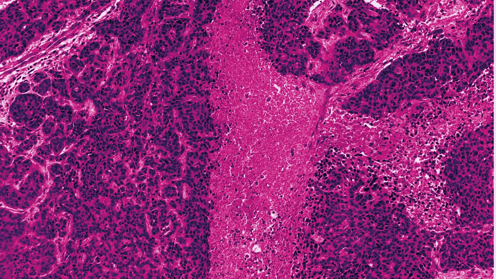
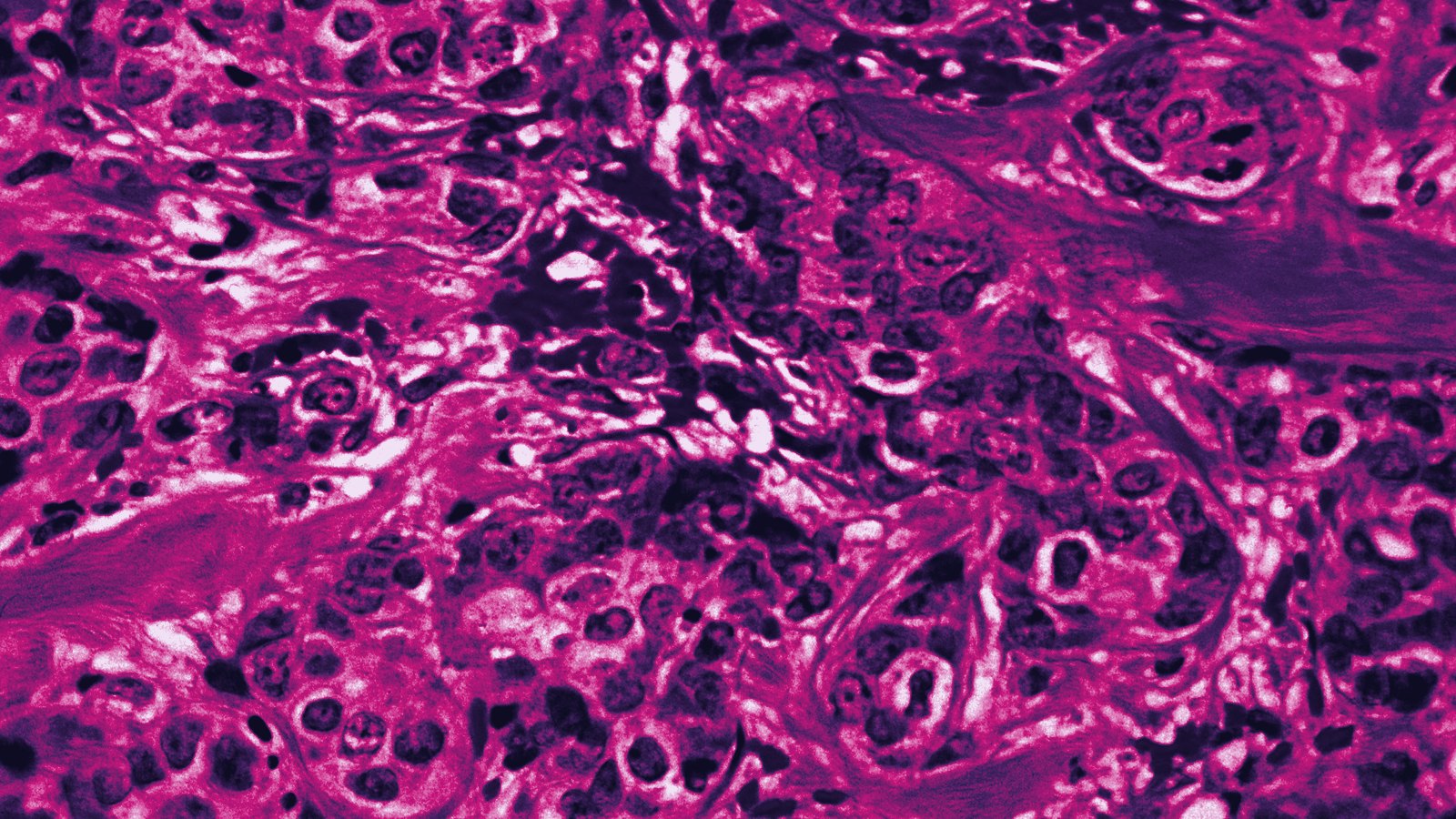
A 711-patient analysis of the randomized SweDCIS trial, published August 24 in JNCI, reports that the proportion of DCIS ducts surrounded by myxoid stroma on the original diagnostic H&E section tracks with later invasive recurrence. Each 10-point increase in that proportion carried an adjusted hazard ratio of 1.16 (95% CI 1.04 to 1.28), and 20-year invasive recurrence was 15.7% for lesions with at least 10% myxoid stroma against 7.1% below that cut-off.
- Cohort: 711 SweDCIS patients treated with breast-conserving surgery, with or without radiotherapy.
- Events: 80 ipsilateral invasive recurrences as first event after surgery.
- Effect size: adjusted HR 1.16 per 10% increase in myxoid stroma (1.04 to 1.28; p = 0.005), cause-specific Cox regression.
- Groups: 296 lesions scored sclerotic (41.6%) and 415 with at least 10% myxoid stroma (58.4%).
- Invasive recurrence: 10.1% vs 4.7% at 10 years and 15.7% vs 7.1% at 20 years for the myxoid and sclerotic groups.
- Authors: Uppsala University, University of Gothenburg, University of Bergen and PreludeDx; JNCI doi 10.1093/jnci/djag293.
A score read from the slide that is already on file
The study used the hematoxylin-eosin sections cut at diagnosis, with no additional stain, and asked two raters to score what fraction of the DCIS ducts sat inside myxoid stroma. Aglaia Schiza and Viktoria Thurfjell of Uppsala University's Department of Immunology, Genetics and Pathology share first authorship, and Fredrik Warnberg of the University of Gothenburg and Carina Strell of Uppsala and the Centre for Cancer Biomarkers at the University of Bergen share the senior credit. The patients came from SweDCIS, the Swedish randomized trial of radiotherapy after breast-conserving surgery for DCIS, which gives the analysis long follow-up and a balanced radiotherapy split of 361 irradiated and 350 non-irradiated women.
In cause-specific Cox regression, invasive recurrence risk rose with the amount of myxoid stroma, and the abstract reports the adjusted estimate as 1.16 per 10% increase. The authors write that "periductal myxoid stroma may be a simple histologic marker for the invasive potential of DCIS," a claim pitched at the overtreatment problem their opening sentence names: clinicopathologic criteria, in their words, "are insufficient to define the invasive potential."
Younger women and larger lesions in the myxoid group
Table S1 shows that the myxoid lesions were not a random slice of the cohort. Women under 50 made up 27.2% of the myxoid group and 19.2% of the sclerotic group (p = 0.002), and lesions over 15 mm accounted for 35.1% versus 23.7%. HER2 3+ status ran 41.1% against 16.3%, nuclear grade 3 ran 48.4% against 23.6%, Ki67 of 20% or more ran 38.4% against 21.7%, and a tumor-infiltrating lymphocyte score of 10 or more ran 51.8% against 18.9%, each at p below 0.001. Screen detection (79.4% overall), margin status, focality and diagnosis year did not differ between the groups, and only 22 women in the whole cohort received endocrine therapy.
Table S2 separates the two kinds of recurrence, and the in situ curves barely move: 20-year in situ recurrence was 13.0% in the sclerotic group and 13.4% in the myxoid group, a difference of less than half a point. The invasive curves are where the groups part: 7.1% and 15.7%, an absolute gap of 8.6 points. Raising the threshold to 33% myxoid stroma leaves 123 patients above the line and narrows the gap to 5.4 points (16.7% versus 11.3%), so the 10% cut-off carries the stronger signal in this cohort.
Main text still unread at press time
JNCI received the manuscript on July 6, accepted it on August 13 and posted it on August 24 as an open-access accepted manuscript available only as a PDF, and that PDF could not be retrieved by APO|APE News at press time. This account therefore rests on the abstract, the author listing and the two supplementary tables, so the covariates in the adjusted model, any inter-rater agreement statistic and any test for interaction with radiotherapy are not reported here. Troy Bremer of PreludeDx in Laguna Hills, California, is a co-author, and the paper's disclosure section was likewise not readable. The patient set is also old, with 69.2% of the women diagnosed in 1995 or earlier.
Why This Matters to the APO|APE Reader
The 8.6-point absolute difference in 20-year invasive recurrence comes from a feature a breast pathologist can add to the report without a new block, a new stain or a send-out, which puts it in a different cost class from gene-expression DCIS assays. Whether it survives adjustment for HER2, grade and TILs is the question the full paper has to answer, because Table S1 shows all three travelling with the myxoid phenotype. If it does, a 10% threshold scored on the diagnostic slide would be the cheapest triage step yet proposed for the 80 of 711 SweDCIS patients whose DCIS came back as invasive disease.
Sources
- Schiza A, Thurfjell V, Holmberg E, Bremer T, Micke P, Karlsson P, Warnberg F, Strell C. Periductal myxoid stroma identifies ductal carcinoma in situ at risk of invasive recurrence. JNCI: Journal of the National Cancer Institute, djag293, published online August 24, 2026
- PubMed record 42635524, abstract and author affiliations. National Library of Medicine, August 2026